Sie befinden sich hier
Inhalt
Date: 13.04.2026
Scientists track individual synapses in the brain over two weeks and show that stronger synaptic connections help stabilize brain structures
A new study by the Center for Molecular Neurobiology Hamburg (ZMNH) and the Mannheim Center for Translational Neuroscience (MCTN) at the Medical Faculty Mannheim of Heidelberg University sheds light on one of the most persistent mysteries in neuroscience: How can the brain maintain stable memories when the physical structures underlying them are constantly changing? The findings by Dr. Cynthia Rais (ZMNH) and Professor Dr. Simon Wiegert, who heads the Department of Neurophysiology at the MCTN, show that the functional strength of a synaptic connection directly predicts how long that connection will persist.
The brain stores memories in distributed networks of nerve cells (neurons) that are connected via synapses. Each neuron has thousands of these connection points ‒ many of them located on microscopic structures known as dendritic spines. Much like transistors in a computer, synapses regulate the flow of information in neural networks. However, unlike computer circuits, which operate in binary states (on/off), synapses are highly dynamic: they can continuously change their strength and may even disappear and reappear over the course of days or weeks. Despite this high level of dynamism, memories can last a lifetime. How the brain resolves this apparent contradiction has remained largely unclear.
“Our results suggest that the brain has an elegant solution to the dilemma between stability and plasticity. What had previously only been hypothesized in many studies, we were able to demonstrate in the living brain: strong synaptic connections are preferentially preserved, while weaker ones are eliminated more quickly,” explains Simon Wiegert. “Moreover, the combined effect of many synapses contributes to the stability of information ‒ a mechanism that may be crucial for long-term memory storage.”
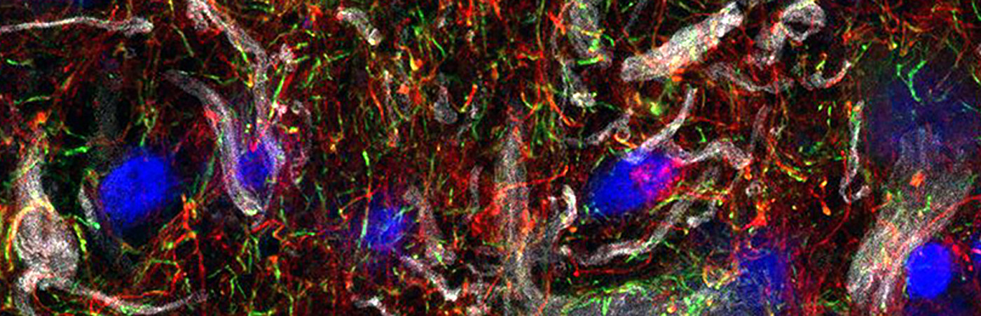
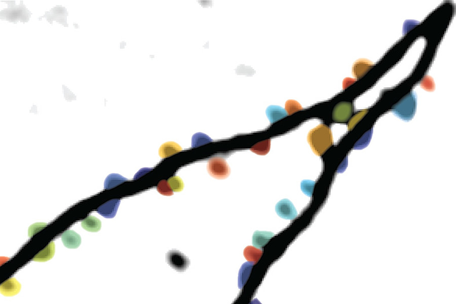
Cynthia Rais and Simon Wiegert used optogenetics in combination with two-photon calcium imaging to track the same dendritic spines in the hippocampus ‒ a brain region essential for many forms of memory ‒ in awake mice over a period of two weeks. They stimulated a specific population of neurons (CA3) and observed how connected neurons (CA1) responded, down to the level of individual dendritic spines.
A particular challenge was that synapses are extremely small ‒ close to the resolution limit of optical microscopes ‒ and movement of the animal often leads to loss of focus. Researchers frequently use anesthetics to avoid motion artifacts; however, anesthetics also alter synaptic transmission and stability, making it difficult to study both function and morphology in the same experiment. Rais and Wiegert overcame this challenge by training the mice to remain still during imaging. Using a closed-loop system, they were able to measure synaptic activity and morphology without any movement-related interference.
This approach allowed them to establish, for the first time, a direct link between the functional strength of dendritic spines and their fate in the living brain. High neuronal activity correlates with stable synaptic connections. Furthermore, the researchers were also able to confirm for the first time in vivo that larger synapses are not only more stable than smaller ones, but also functionally stronger.
Another key finding of the study was that, despite the high variability of individual synapses over time, signal transmission at the level of dendritic branches remained constant. In other words, the brain maintains a reliable flow of information at this level. This stability of information transfer was also observed at the level of entire neurons, which showed consistent responses to the same stimuli across multiple sessions.
Thus, a high degree of redundancy in synaptic information flow can stabilize communication between neurons. This observation may help resolve the puzzle of how the brain preserves stable memories despite ongoing dynamic changes in its fundamental communication units ‒the synapses. It is similar to an ant trail: individual ants may move unpredictably, but the colony as a whole maintains a stable path.
The present study helps improve our understanding of the mechanisms underlying neurodegenerative diseases such as Alzheimer’s, a key feature of which is the early loss of synapses. At the same time, it raises new questions about what happens to this stabilization process during learning and memory formation, when synapses are selectively strengthened or weakened. These processes, known as long-term potentiation and long-term depression, are considered fundamental to the formation and updating of memories.
Publication:
Rais C, Wiegert JS.
Functional synaptic connectivity shapes spine stability in the hippocampus.
Nature Communications 17(1):3218 (2026).
DOI: https://doi.org/10.1038/s41467-026-71332-z
Press release of the Medical Faculty in Mannheim (in German):
09.04.2026 Neue Erkenntnisse aus der Gedächtnisforschung